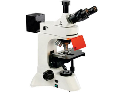
A fluorescent microscope is a type of microscope that uses fluorescence and phosphorescence to create an image. It uses a beam of light, usually ultraviolet, to excite fluorescent molecules in the sample, causing them to emit light at a different wavelength (usually visible light). This emitted light is then captured by the microscope's optics and used to create an image of the sample. Fluorescent microscopy is commonly used in biological research, as it allows for the visualization of specific molecules or structures within cells.
Application of fluorescent microscope
Fluorescent microscopy has a wide range of applications in biology and medicine, including:
Cell biology: Fluorescent dyes can be used to label specific cellular structures, such as the nuclei, cytoskeleton, or mitochondria, allowing for detailed analysis of cell structure and function.
Immunofluorescence: Antibodies that have been conjugated to fluorescent dyes can be used to specifically bind to and label proteins of interest in cells or tissues.
Microscopy of live cells: Fluorescent dyes that do not damage cells can be used to label live cells, allowing for the study of cellular processes in real-time.
Genetics and molecular biology: Fluorescent dyes can be used to label DNA and RNA, allowing for the study of gene expression and regulation.
Pathology: Fluorescence-based techniques are used to detect cancer cells or bacteria in tissue samples,
Drug discovery: Fluorescence-based assays can be used to measure the activity of drugs and drug candidates on specific cellular targets.
Material Science: Fluorescence microscopy is also used to study the properties of materials and particles, such as the size, shape and composition of nanoparticles.
Environmental Science: Fluorescence microscopy can be used to detect and quantify microorganisms in water and soil samples.
